1 of 7 / Overview
View All
What Is Substance Abuse?
Substance abuse differs from addiction. Many people with substance abuse problems are able to quit or change their behavior.
What Is Drug Addiction?
Addiction is a disease that affects your brain and behavior. With drug addiction, you can’t resist the urge to use them.
Signs of Drug Addiction
If you’re worried that you or a loved one may have an addiction, there are signs to help you know.
Tolerance, Physical Dependence, and Addiction Explained
"Addiction,” “tolerance,” and “physical dependence” are often used interchangeably. But they don’t mean the same thing.
2 of 7 / Alcohol Use Disorder
View All
What Is Alcohol Use Disorder?
Alcohol use disorder (AUD) is a chronic illness in which you can’t stop or control your drinking.
Alcohol Use Disorder: Myths and Facts
Are you falling for common misconceptions about alcohol use disorder?
The Stages of Alcoholism
No one becomes addicted to alcohol overnight. You don’t catch alcoholism the way you do the common cold.
Do I Have an Alcohol Problem?
If you've had two or three of these symptoms in the past year, that’s a mild alcohol use disorder.
3 of 7 / Opioid Use Disorder
View All
Painkillers and Opioid Use Disorder
A national health crisis, 3 million people in the U.S. have opioid use disorder (OUD), or they’ve had it in the past.
Opioid Abuse Statistics: Who’s Affected and Why?
According to the National Center for Drug Abuse Statistics, 3.8 percent of American adults abuse opioids each year.
Fentanyl: What You Need to Know
Fentanyl is a human-made opioid used to treat severe pain. It's 50 to 100 times stronger than morphine.
Heroin
Heroin is a drug that comes from a flower, the opium poppy, which usually grows in Mexico, Asia, and South America.
4 of 7 / Cannabis Use Disorder
View All
What Is Marijuana Abuse?
Marijuana misuse can have dire consequences if left untreated. It’s possible to misuse it and get addicted to it.
Recreational Marijuana FAQ
Despite increasing acceptance, marijuana use does have some risks. Here's a look at how marijuana can affect your health.
How Pot Affects Your Mind and Body
Marijuana has mind-altering compounds that affect both your brain and body. It can be addictive, and it may be harmful.
How to Avoid a High Tolerance to Cannabis
Long-term use of cannabis can reduce the effects of its intoxication. So you can develop a tolerance for the drug.
5 of 7 / Prescription Drug Abuse
View All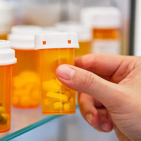
What Is Prescription Drug Abuse?
Prescription drug abuse is when you take a medication for a reason other than why the doctor prescribed it.
Prescription Addiction: Who's at Risk?
Addiction is a complex disease brought on by many reasons, including your lifestyle and genes.
Should You Worry About Your Back Pain Meds?
Opioid meds do relieve back pain for short periods, but they're strong -- and they come with some serious risks.
Dealing with Side Effects of Percocet
Percocet is a mix of acetaminophen and oxycodone. Taking it doesn't come without side effects.
6 of 7 / Other Abused Substances
View AllStreet Drugs: Know the Facts and Risks
With street or club drugs use, you’re taking risks. The drugs are dangerous, and there’s no way to know how strong they are.
What Is Cocaine?
Cocaine is a highly addictive drug that ups your levels of alertness, attention, and energy.
Crystal Meth: What You Should Know
Crystal meth is the common name for crystal methamphetamine, a strong and highly addictive drug.
What Is Kratom?
Doctors believe some substances in kratom attach themselves to the same parts of a nerve cell as opioid painkillers.
7 of 7 / Non-Substance Addictions
View AllSigns of a Sex Addict
Sex addiction is defined as a lack of control over sexual thoughts, urges, and impulses.
Porn Addiction
Porn addiction is, in theory, when you can’t stop looking at porn, even if you want to.
What Is Overeaters Anonymous?
Overeaters Anonymous are groups of individuals who come together to meet over a shared problem: compulsive overeating.
Suggested Reads about Substance Abuse and Addiction
Studies: Smoking Pot Affects Driving Longer Than People Think
Studies conducted by the Center for Medicinal Cannabis Research at the University of California, San Diego, show people should wait about 4.5 hours after smoking one joint before they attempt to drive a car,
Lab Testing Detects Synthetic Pot in ‘Gas Station Heroin’
A new CDC report says synthetic cannabis has been found in some “gas station heroin” products, which are often marketed as mood-boosting supplements that can help with alertness or energy.
Rise in Overdose Deaths Driven by Drug Combinations
Reports of the use of fentanyl along with the animal tranquilizer xylazine makes preventing substance use disorder and associated overdose deaths even more complicated.
FDA Approves First Test to ID Potential Risk of Opioid Addiction
The FDA has approved the first test that uses DNA to assess if people might have a higher risk of becoming addicted to opioids.
Top Search Terms for Substance Abuse and Addiction
8 million+ Physician Ratings & Reviews
Find Doctors and Dentists Near You
You can also search by physician, practice, or hospital name